What are peripheral and carotid procedures?
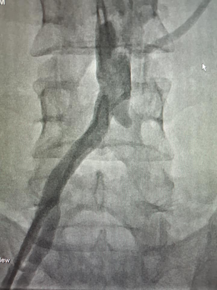
Peripheral angiography examines arteries outside the heart, including vessels supplying the legs. Carotid angiography examines arteries in the neck that supply the brain. Imaging can help define the location and extent of disease.
Angioplasty uses a balloon to widen a selected narrowed segment. A stent may be placed to support the artery. Treatment is chosen after reviewing symptoms, anatomy, other investigations and alternatives.
When might a procedure be considered?
Peripheral artery disease may cause walking-related leg symptoms, poor wound healing or other evidence of impaired circulation. Carotid disease requires a separate assessment of stroke history, symptoms and overall stroke risk.
An imaging finding alone does not decide treatment. Medical therapy and risk-factor management remain important, and some people are better suited to surgery or another approach.
What happens during angioplasty or stenting?
A catheter is introduced through a blood vessel, commonly at the groin or sometimes the arm. Contrast and X-ray guidance help the operator assess the artery. A wire, balloon and, when appropriate, a stent are used to treat the selected narrowing.
Carotid treatment needs particular attention to the risk of material travelling towards the brain. The specialist team explains the planned technique and individual risks before treatment.
Recovery and longer-term care
Monitoring includes the puncture site and the circulation or neurological status relevant to the procedure. Follow-up may include vascular imaging and a review of symptoms, medicines and risk factors.
Risks include bleeding, vessel damage, contrast reactions, kidney problems and recurrence of narrowing. Carotid stenting also carries a stroke risk. The expected benefit and alternatives should be discussed for your individual situation.
Frequently asked questions
Is every narrowed carotid artery treated with a stent?
No. The choice may include medical treatment, carotid surgery or stenting, depending on symptoms, narrowing, anatomy and individual procedural risk.
Are the before-and-after images a prediction of my result?
No. They are labelled clinical examples from the supplied archive. Angiographic views can differ, and an individual outcome cannot be predicted from another patient’s images.
Does a stent replace ongoing treatment?
No. Follow-up, prescribed medicines and management of vascular risk factors remain part of care after a procedure.